SB-4982
41 y/o female with a long-standing history of "chronic sinusitis" resistant to medical therapy.
Endoscopy/CT revealed pansinusitis, sphenoid and ethmoid "polyps," and opacification of her superior nasal cavity and mucosa. Gross examination revealed multiple irregularly-shaped fragments of soft pale gray tan to reddish-brown tissue, 0.2 cm to 1.5 cm.
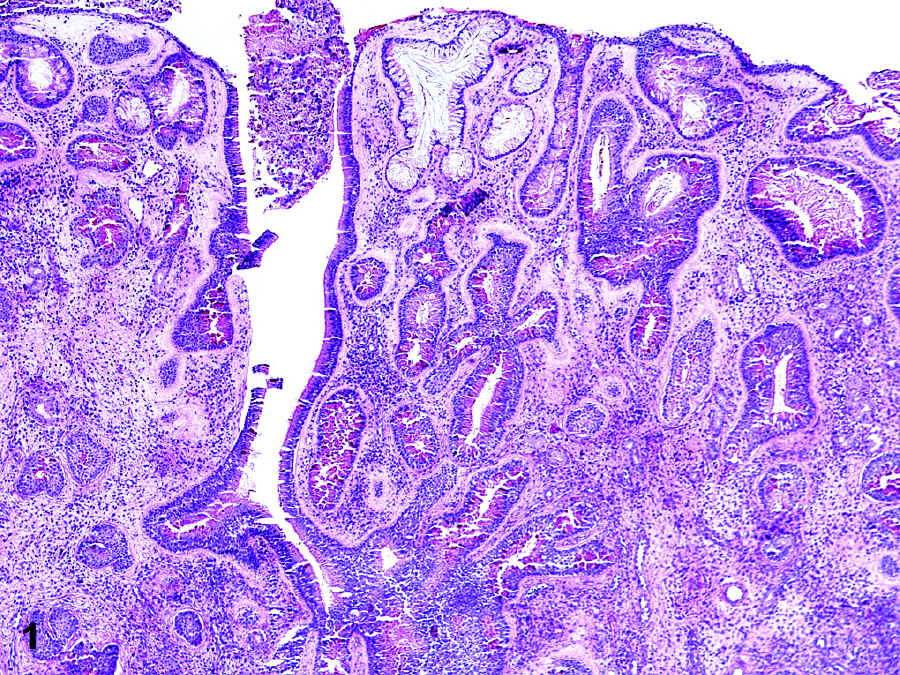
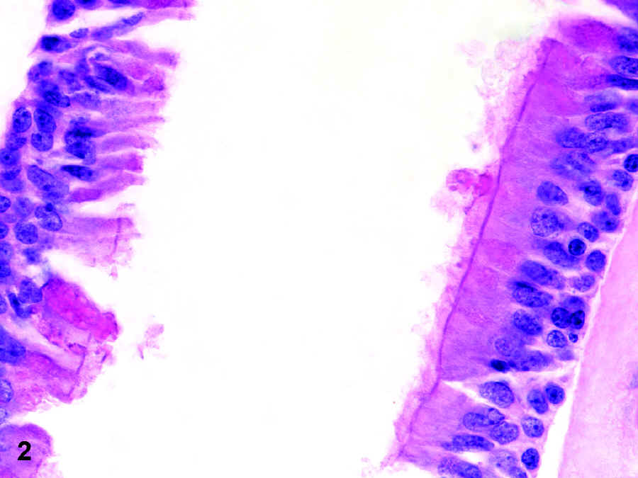
MICRO: Figures 3-5.
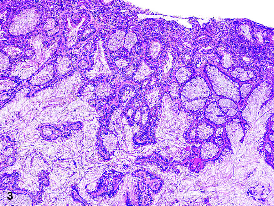
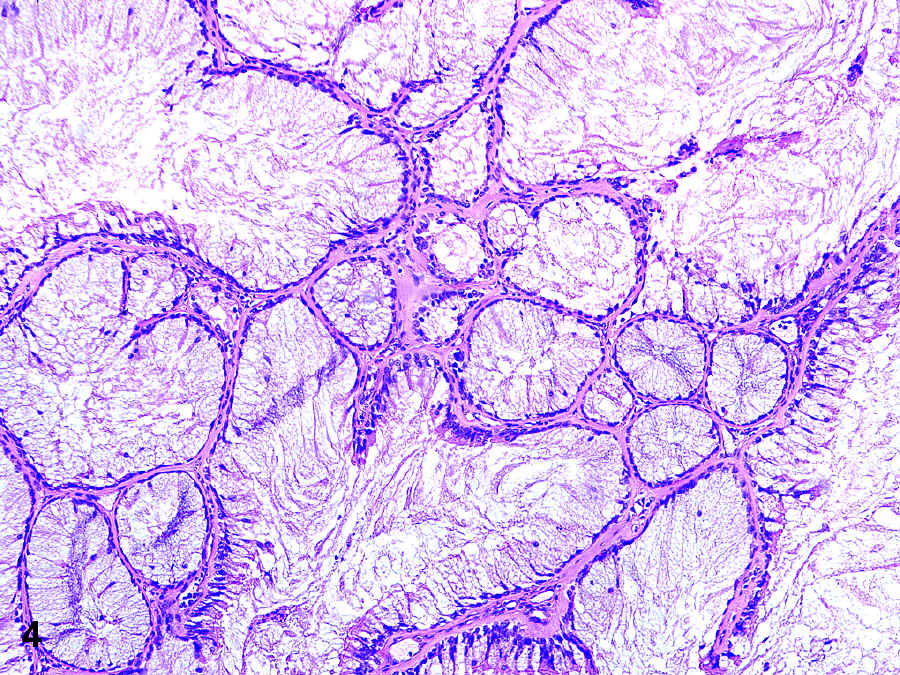
Subsequent MRI showed moderate soft tissue enhancement of the maxillary sinus; additional paranasal sinus biopsy revealed Figures 1-2.


